
Published on the 5th April 2018 by ANSTO Staff
The complementary power of combining multiple X-ray techniques to understand the unusual properties of hyperaccumulator plants has been highlighted in a new cover article just published in New Phytologist.
Among the different X-ray techniques that were evaluated in the paper, each has its own advantages. Kopittke said he was a strong proponent of XFM for this very reason.Elemental distribution maps (Ni, Co, Ca) obtained with X‐ray fluorescence microscopy (XFM) (left) and scanning electron microscope (SEM) (right) of an identical sample (Rinorea javanica leaf).
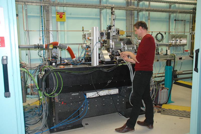
XFM beamline scientist Dr Martin de Jonge, a co-author on the paper with Dr David Paterson, said the technique was used to study how metals are taken up in roots and distributed throughout the plant under ‘lifelike’ conditions.
“Because a vacuum is not required, you can undertake the experiment in ambient conditions. Plants are more than 90 per cent water and if you have to dehydrate them to put them in a vacuum as you do for other X-ray techniques, you invariably move the elements you are analysing. With XFM we can analyse a live plant in its natural state, and this is highly advantageous. You cannot do this with any other technique, “said Kopittke.
The authors note that as XFM can probe to a depth from several hundred micrometres to the millimetre scale, it is ideal for addressing plant science. “Its high sensitivity and capacity to be used on large samples make it very versatile,” said de Jonge.
Unique to the XFM beamline is the use of a revolutionary X-ray detector Maia which is an extremely fast detector system. Maia makes it possible to obtain elemental maps that are many millions of pixels (megapixels) in size in hours. Because of this, there was no visible evidence of radiation-induced damage to live plants.
XFM can simultaneously map hyperaccumulated metals (such as nickel), important nutritional elements (such as potassium) and trace elements that are typically present at far lower concentrations (such as copper).
“You can illuminate with a single energy and you get the fluorescence from everything, known as the spectral axis,” said de Jonge. “But you can also modify the incident energy and look at changes in the spectral axis as a result of that.This helps you probe the electronic structure of atoms.“
The other techniques that were comprehensively reviewed in the paper included proton induced X-ray emission (PIXE). This technique can be undertaken on frozen samples; thereby making it possible to investigate cross-sections of plant tissue to elucidate the internal cellular-level distribution of elements. PIXE is also ideally suited to measure very light elements, such as aluminium.
“The complementarity of X-ray elemental mapping techniques provides a way to answering questions at every level about how plants take up metals and maintain their physiological functions” said Kopittke.
Collaborating institutions included The University of Queensland, University of Adelaide, Université de Lorraine (France), iTHEMBA Labs (South Africa), University of Science and Technology (Poland), Jagiellonian University (Poland), and CSIRO.
https://doi.org/10.1111/nph.14810