
Senior Principal Research Scientist

Showing 1 - 20 of 23 results
ANSTO is participating in a new Australian Research Council (ARC) Centre of Excellence for Indigenous and Environmental Histories and Futures (CIEHF) to be headquartered at James Cook University (JCU) that aims to bring Indigenous and environmental histories to the forefront of land and sea management.
A new radiocarbon dating facility opened at the University of New South Wales (UNSW) will complement the extensive radiocarbon facilities at ANSTO’s world-leading Centre for Accelerator Science
Stronger research link with IAEA with the establishment of Collaborating Centre at ANSTO to support environment and cultural heritage activities


Dr Karina Meredith was appointed Director of the new Research and Technology Group for Environment effective 15 January 2024.
Insights about Mayan Empire relevant for current climate challenges

Since 1962, the United Uranium Scholarship has helped promising young scientists in the field of nuclear energy extend their knowledge and expertise. In 2022, scholarships were awarded to several ANSTO researchers, including Phil Sutton.

Particle induced X-ray emission can be used for quantitative analysis in archaeology, geology, biology, materials science and environmental pollution.
Read about an ANSTO scientist and their work to prepare for a school project or interview.
Particle Induced X-ray Emission (PIXE) is a powerful and relatively simple analytical technique that can be used to identify and quantify trace elements typically ranging from aluminium to to uranium.

ANSTO User Meeting 2021 - Speakers
Young and mid-career ANSTO scientists and engineers have been featured in the latest issue of Careers with STEM that highlights careers in nuclear science.
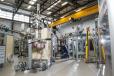
The BRIGHT Project will expand the beamline infrastructure of the Australian Synchrotron to increase both its capacity and capabilities.
A rare collection of traditional Aboriginal wooden objects in varying degrees of preservation found along a dry creek bed in South Australia have been dated to a period spanning 1650 to 1830 at the Centre for Accelerator Science at ANSTO.
The Medium Energy- X-ray Absorption Spectroscopy beamlines provides access to XANES and EXAFS data from a bending magnet source, optimised for cutting-edge applications in biological, agricultural and environmental science in an energy range that is not currently available at the Australia Synchrotron.