

Published on the 9th November 2016 by ANSTO Staff
Early career environmental researcher, Divya Vinod has used radioactive isotopes at ANSTO to investigate selenium uptake in plants to assess their suitability for use in remediating contaminated environments, such as mining sites.
|
| Vitamin greens and sunflowers were investigated for their ability to absorb selenium |
Although selenium is an essential element for humans and animals, elevated levels of selenium in soil and water found near coal, phosphate, uranium and some metal mines can be toxic to human health, affect agricultural productivity and the stability of natural ecosystems.
Under the supervision of ANSTO Ecotoxicologist Dr Tom Cresswell, Vinod has used whole-plant imaging of radioactive selenite and selenate to determine where the selenium is accumulating in two edible plants, vitamin greens (Brassica rapa cv.) and dwarf sunflowers (Helianthus annus cv.) amongst others to determine how they accumulate selenium.
Hyperaccumulator plants thrive in soils with high concentrations of metals which they absorb through their roots.
In addition to winning ‘Best Presentation’ at the Young Researcher Mini Conference at ANSTO in August, Divya received the Best Student Oral Presentation Award at the Society of Environmental Toxicology and Chemistry (SETAC) Australasia 2016 Conference in October.
“By adding radioactive isotopes to a controlled system in the laboratory, it is possible to follow how a particular element behaves and how it is bioaccumulated by the plant or organism,” explained Cresswell, who uses the techniques to investigate contaminant bioaccumulation in marine organisms.
Selenium was bombarded with neutrons to create the radioisotope, 75Se which gives off radioactive signals that can be rapidly detected.
The 75Se as selenite and selenate was added to a solution, in which vitamin greens and sunflower plants were growing.
Selenite and selenate are the two forms of selenium that are taken up by plants.
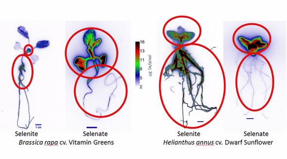 |
| Radiographic images depict where the concentrations of selenium are found |
After 21 days of exposure to pre-determined concentrations of both selenite and selenate salts, Vinod took radiographic images of the plants, where the radioactive selenium was clearly visible (depicted above).
The imaging allowed Vinod to make some semi-quantified assessment of biodistribution of the selenium.
In both plants, the greater concentrations of selenite occurred in the roots, while more selenate accumulated in the leaves.
Vinod also used protein fractionation techniques in the research, which suggests the selenium is binding to albumin, crude starch and glutelin proteins.
Hyperaccumulation of selenium in plants is not completely understood but this group of plants possess a unique physiological characteristic that may enable plants to prevent selenium from poisoning plant cells by disrupting biochemical reactions and specific enzyme functions.
She intends to do further proteomic studies at the University of Technology Sydney under the direction of Dr. Luigi DeFilippis to confirm if selenium is being incorporated into a specific protein class which may provide information on plant protein function.
Vinod is hopeful that a better understanding of selenium accumulation, and metabolism is likely to have significant implications for determining the plant’s suitability for phytoremediation.
The Australian Institute of Nuclear Science and Engineering (AINSE) provided partial funding to facilitate the research.