

Showing 1 - 10 of 10 results
OPAL reactor back online after planned long shutdown
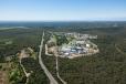
ANSTO’s OPAL multi-purpose research reactor at Lucas Heights has officially returned to power and recommenced operations, following a months-long planned shutdown to carry out essential maintenance and upgrades.
Statement of Intent
In January 2026, Mr Michael Quigley AM, ANSTO Board Chair, shared ANSTO's Statement of Intent with the Minister for Industry and Innovation, and Minister for Science.
Licence awarded
Australia’s new state-of-the-art nuclear medicine facility gets green light.
Nuclear Medicine
ANSTO manufacture and supply a range of radiopharmaceuticals, radiochemicals, kits and accessories for use in research, industry and the health sector.
Bringing radiochemistry to life
ANSTO to undertake routine shipment of spent fuel in 2025
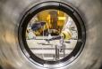
Innovative radioactive waste treatment technology forging ahead
International interest is building in Australia’s new multi-million-dollar radioactive waste processing facility at the Sydney campus of the Australian Nuclear Science and Technology Organisation (ANSTO).
Exceptional group of women at the forefront of science outreach at ANSTO
Five exceptional female science communicators are part of a larger team who use skills in education and engagement to promote an interest in science amongst the public and students.
Samples - Infrared microspectroscopy
The Infrared microspectroscopy microscopes can record spectra from a range of different samples; from thin microtomed sections to polished blocks and embedded particles. This section highlights the types of samples that can be analysed using the IRM beamline