Experiments using an advanced imaging technique at ANSTO’s Australian Synchrotron have clarified how pink porous volcanic rock, found in the ocean following an eruption, might be formed in an underwater explosion.
Researchers from the University of Louisiana Lafayette, the Australian National University, and Queensland University of Technology applied X-ray fluorescence microscopy on samples of pink and white pumice found floating following the 2012 eruption of Havre Seamount, an undersea volcano in the Southwest Pacific Ocean.
The investigations clarified the source of pink colouration by examining the proportion and distribution of magnetite and hematite in the pumice samples.
In research published in Communications Earth and Environment, they concluded that the pink colourisation is due to the oxidation of magnetic nanolites and microlites to haematite and that a substantial proportion of magnetic microlites and nanolites need to be available for this to occur.
The Lafayette University researchers suggested that the findings challenge the known depth limits for deep-sea eruptions and assumptions that deep-sea eruptions can’t be explosive.
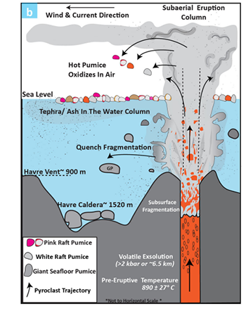
The pink colourisation supported the theory that it was launched by a powerful jet from more than a mile beneath the ocean floor at temperatures above 700 degrees Celsius (1,300 degrees Fahrenheit). Read more on the Lafayette University website and a report on the QUT website.
The pumice becomes pink when tiny magnetic microlites and nanolites are exposed to extreme temperatures of magma when the hot pumice in the explosive eruption column reaches the atmosphere and oxidation takes place.
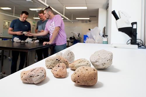
Both white and pink pumice are produced in eruptions and form a raft on the ocean’s surface.
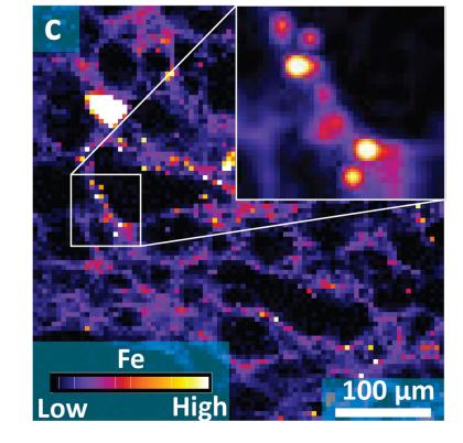
“The spectroscopy confirmed that there is more magnetite and haematite in pink pumice than white pumice,” said Senior Instrument Scientist Dr Daryl Howard, a co-author of the publication.
“The iron-based magnetite and haematite can be detected and measured on our beamline,” he added.
Seafloor pumice, which is white, contains only magnetite. However, white raft pumice contains magnetite and small amounts of haematite.
The recent eruption of another undersea volcano, Hunga Tonga-Hunga Ha’apai, made headlines in March this year with a blast that is believed to be the loudest terrestrial event since Krakatoa in 1883 and was captured by space satellites.
In contrast, the eruption of Havre Seamount was largely unnoticed until the raft pumice was discovered.
Dr Eric Ferré of the University of Louisiana undertook research on the magnetic properties of the pumice, in collaboration with one of his former students, Joe Knafelc, now at Queensland University of Technology (QUT), in Brisbane and Dr Scott Bryan and Dr Michael Jones, also at QUT.