
The cryogenic electron microscope (Cryo-EM), commissioned and operated by the University of Wollongong (UOW), supported by a number of collaborating institutions and temporarily housed at ANSTO’s advanced microscopy facility, provides an extraordinary ‘window into the cell’ that is leading to important insights into neurological disorders, other diseases, drug discovery, and basic science.
The powerfully-engineered Titan Krios is one of only two operating in Australia. It will ultimately be housed in the UOW’s purpose-designed Molecular Horizons Building when it is constructed.
The development of the technique was so significant it earned the three scientists who pioneered it the Nobel Prize in Chemistry in 2017.
“This instrument is accelerating advances in structural biology and to have this capability based at ANSTO for our scientists and other users is a real privilege. Working in a true partnership with UoW we provide operational support at our end which complements the remote operation from the Northfields Avenue campus of the university,” said Dr Miles Apperley.
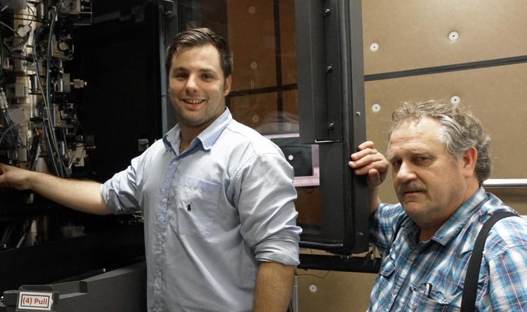
At ANSTO, Dr Daniel Oldfield and Ken Short have assisted with a range of technical services to support the daily operation of the instrument.
“ANSTO’s advanced microscopy building has the advanced features needed to shield the sensitive instrument from vibrations as well as magnetic and electric fields,” said Oldfield.
Scientists use the technology most often to study the structure and function of important biological building blocks, such as proteins, which are comprised of sequences of amino acids. They are typically between 3-10 nanometres in size.
A nanometre is a billionth of a metre and a human hair is 75,000 nanometres in width. The ultimate resolution of 3D structures can be determined with a Krios, which resolves on the order of 1.5 angstrom. This is sufficient to locate atoms within a protein structure (an angstrom is a 10th of one nanometre).
Physics World has reported that the technique’s particular strength is its ability to construct models of single, unattached molecules from images of the molecules in their natural state – encompassing a range of arrangements and binding states with other molecules.
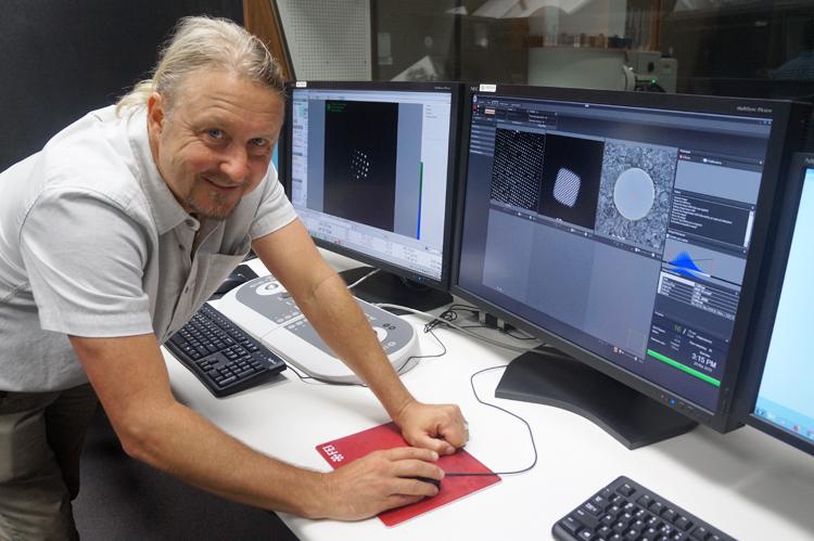
“The structure of these complex macromolecules can be captured at an atomic resolution, which is equivalent to seeing where every atom in the molecule is located,” explained Dr James Bouwer (pictured above), General Manager of Cryogenic-electron Microscopy in the University of Wollongong’s Molecular Horizons Program, who is a pioneer in its use.
“The instrument’s ability to produce 3D maps of key structures, allows you to see what cannot be determined using other techniques, such as X-ray crystallography or nuclear magnetic resonance.”
The technique involves rapid cooling of a solution containing a protein or other molecule until it forms an amorphous or glass ice. Extremely thin samples of only a few 100 nanometres are excised, refined using focussed ion beam milling and then placed in the microscope where they are probed by a stream of electrons.
A direct detector can count every electron that strikes. The advanced detectors have ultra-sensitive camera systems that collect images of individual single particles in different orientations.
“Wherever a single particle freezes, it will occur in a specific orientation randomly. Particles in the same orientation in two dimensions can be grouped and classified for further refinement using powerful computations,” explained Bouwer.
Software is used to process the images, correct motion, select particle images in random orientations and classify 2D images. The 2D projections can be used to reconstruct 3D images.
“We need high-speed networks and the computing power of Monash’s MASSIVE supercomputer to do this,” said Bouwer.
Refinements are necessary because the signal to noise ratio of averaged projections increase dramatically.
The instrument generates about two terabytes of data a day.
Bouwer emphasises that the operation of the technology involves support from mathematicians, computer scientists, physicists, chemists, biologists, engineers, and imaging experts.
The low-dose technique has superior advantages over conventional electron microscopy, where the high energy of the electrons destroys a sample quickly.
Because a lower electron dose is distributed between many images of copies of the same protein particle using the cryo-electron microscope, thousands of noisy images can be summed together to create high ‘signal-to-noise’ images of the protein of interest.
The primary advantage over X-ray crystallography is that the technique does not require solid crystals of a molecule.
The samples can be imaged in their natural environment, which is typically an aqueous solution.
The approach is also fast compared to the time consuming, laborious staining techniques that preceded it in the decades before its introduction.
“We can put a sample on and have a structure the next day,“ said Bouwer.
However, Bouwer says that it was the development of direct electron detectors, something that he contributed to at the University of Southern California, San Diego at the National Center for Microscopy and Imaging Research under Mark Ellisman that brought a revolution in CryoEM.
“The new detectors can count every electron that strikes the sample, which delivers a strong signal,” said Bouwer.
“We can say with certainty if an electron hit or it didn’t the detector. A high-speed readout of up to 400 frames/second allowed us to correct for sample drift during imaging. This allows us to see the atoms, rather than having them smeared across a couple of second images.”
Crystallographic electron density maps created using X-rays are often docked with the cryo-electron microscopy models of the same molecule.
“It serves to confirm structure,” said Bouwer.
Another technique, cryo-electron microscopy technique, sub-tomogram averaging, collects data from a series of tilts of nearly identical structures and aligns 2D images to generate an average 3D image.
Bouwer said there was considerable interest from across Australia and the region, as well as collaborations linked to US research.
In Australia, the microscope is being used to look at ion channels in the heart for the Victor Chang Institute.
Researchers at the University of Wollongong is investigating the proteins involved in DNA replication helicases.
Understanding the sequence of amino acids is crucial for drug development.
Before he came to Australia, Bouwer was involved in work at the University of California, San Diego on a protein that bound to microtubules and was involved in Parkinson’s disease.
“Once you get a resolution of 1.8 angstroms or better you can understand where every amino acid in the chain is according to its shape,” said Bouwer.
Investigators who wish to use the cryogenic-electron microscope should refer to information on the University of Wollongong website.