
Basic physical principles
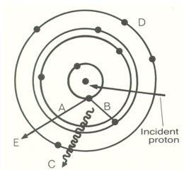
As a charged particle moves through a material, it loses energy primarily by exciting electrons in the atoms that it passes by. Electrons in the inner shells of the atom (predominantly the K and L shells) are given enough energy to cause them to be ejected, resulting in an unstable electron atomic configuration.
The Coulomb interaction between the ion and the target nucleus results in the ejection of an inner shell (K, L etc.) electron (A). The resulting inner shell vacancy is filled by an outer electron (B) and an X‑ray (C) carries off the excess energy. These X‑rays are characteristic of the excited atoms (D)
Electrons from higher shells in the atom then 'drop down' to fill these vacancies, and in so doing, give off excess energy in the form of X-rays. The energies of these X-rays are characteristic of the element and therefore can be used to identify elemental composition. On the other hand, by measuring intensities of characteristic X-ray lines one can determine concentrations of almost all elements in the sample down to approximately 1 ppm (part-per-million).
PIXE technique
PIXE is a powerful and relatively simple analytical technique that can be used to identify and quantify trace elements typically ranging from Al to U. Sample irradiation is usually performed by means of 2-3 MeV protons produced by an accelerator (at ANSTO by STAR and 10 MV tandem accelerators).
X-ray detection is usually done by energy dispersive semiconductor detectors such as Si(Li) or High Purity Germanium detectors. An example of PIXE spectra is shown on the figure below. An example of a PIXE spectrum obtained from an aerosol sample is shown below. This spectrum was accumulated in less than 5 minutes, using 2.6 MeV protons with a 10 nA beam current.
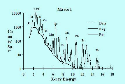
Figure 1: PIXE spectrum of an aerosol sample showing the characteristic
X-ray lines and intensities from the trace elements in the
aerosol particle
Sample preparation: there is virtually no need for sample preparation. Most samples are analysed in their original state; aerosol filter, archaeological samples, soil, biological samples, etc. However, as PIXE technique is probing only top 10 to 50 micrometers of the sample (depending on the material, energy of incident beam and most importantly on the energy of characteristic X-rays), it is very important that the area/volume irradiated by the beam (usually a circular area with the diameter of 1 to 10 mm) is representative of the whole sample.
Therefore, if the sample is obviously not homogeneous, like for example some pottery and geological samples, it is advisable to grind the sample to a fine powder (preferably with particle size smaller than 1-2 micrometers), thoroughly mix it with 20% analytical grade carbon powder and consequently press into pellets. To find more about sample positioning and size visit the Broad Beam Analysis page.
Detection Limits
Sensitivities of PIXE range from 1/100 ppm depending on the element, energy and total charge of incident particles, x-ray filters and the quality of the X-ray detector used.
Applications
The PIXE technique can be applied to a wide variety of sample materials. At ANSTO, PIXE is often used for quantitative analysis in geology, archaeology, biology, materials science and environmental pollution.