
Neutron tomography may soon play a useful role in palaeontological research offering some of the most detailed scans of fossils while dramatically reducing any risk of damage to the specimens. Ada Klinkhamer from the Australian Age of Dinosaurs Museum visited ANSTO to learn more about the technique and shares her experience.
Preparation of fossils that are entombed in rock can be a real nightmare. There might be enough of the fossil exposed to tell what it is and which way is up, but what about the fine details such as delicate antennae on a fossil crayfish, or the intricate neural processes of a small vertebra?
Applying medical and industrial innovations like MRI, CT scanning and, more recently, neutron tomography to palaeontology has made it possible for scientists to delve further into our understanding of the Earth’s past than ever before.
Making fossils visible with the aid of computer technology can help us discover exactly what is in ‘that rock’ without having to prepare it to find out. This minimises greatly the risk of destroying delicate appendages. In fact, computer technology can enable palaeontologists to carry out research on the internal structure of a fossil without having to physically prepare it, or break it open at all.
The Australian Age of Dinosaurs Museum of Natural History (AAOD) research team are always keen to try out these new technologies to see how they might benefit the preparation and research of specimens in their collection. It is because of this interest that I was given the opportunity to visit Sydney for a couple of days to join the team at ANSTO and experience neutron tomography first hand.
Neutron tomography is a relatively new research method that is starting to gain popularity around the world and has only just recently become available in Australia. Its main use is in the aerospace industry and in steel, oil and gas industries where it is used to detect things such as potential faults in propellers, or problems like water damage, or fuel flow in engines. ANSTO houses one of only a few research reactors in the world, and their neutron tomography instrument is called Dingo.
Dingo works by ‘firing’ neutrons at an object through a beam tube from the reactor. Some of the neutrons are absorbed by the object while others pass straight through. A camera on the opposite side of the object is able to capture images of the shadow projected onto a detector as the object is slowly rotated 360o.
These separate 2D images are then fitted together and turned into a 3D image. The resulting image looks much like a CT scan, except CR works by detecting dense material within lighter materials (like bone inside soft tissue), whereas neutron scanning is sensitive both to changes in density and to different minerals in rocks.
Neutron tomography produces a large amount of radiation, meaning the objects being scanned often have an afterglow and need to ‘cool’ for hours, even days, in a secure area until the radiation dissipates. The technique is non-destructive, and the mineral samples can be returned to the museum unchanged.
Our first contact with ANSTO was when AAOD staff member Ben Galea met the Scientific Co-ordinator for ANSTO’s Bragg Institute, Dr Joseph Bevitt, at a National Youth Science Forum in Canberra in 2013.
Ben was sure that Dingo would be useful in identifying fossils in rock at AAOD and arranged for the Museum’s collection Manager, David Elliot, to talk with Joseph. The result was ANSTO kindly offering AAOD ‘beam time’ as part of Dingo's testing and fine-tuning process as they prepare the instrument for academic and commercial projects.
David subsequently packed up and sent them a selection of fossils, including several Crustaceous crabs and crayfish and a couple of pieces of sauropod bone, in the hope that the ANSTO team might be able to reveal what was hidden within the rock.
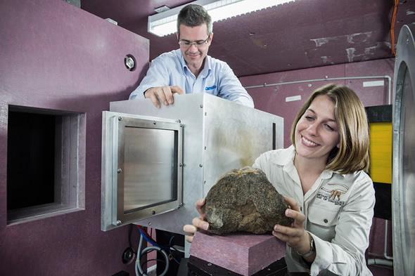
As part of my PhD on Virtual Reconstruction and Biomechanical Analysis of Australian Sauropods, I was thrilled to head to Sydney to meet the team and observe the neutron tomography process first-hand. Talk about an adventure! I arrived just as they were starting work on the AAOD fossils and, after going through security, was met by Joseph and Dr Ulf Garbe who are in charge of the instrument. Entering the Neutron Guide hall was quite an experience.
It was a massive room, the size of an aircraft hangar, and filled with large and complicated looking instruments all named after different Australian animals. Each instrument in the hall can take up to 10 years to build and all utilise nuclear technology in some way to conduct research. Ulf showed me around and explained what all the instruments did, and finally I was introduced to Dingo. For two days I helped the Dingo team scan AAOD fossils with varying degrees of success.
Some of the crayfish and crab specimens came out beautifully; we were able to see full 3D images of the specimens inside their rocky tombs. Unfortunately, scanning the largest pieces of dinosaur bone was unsuccessful. In this case, the specimens were too dense and thick and the neutrons were unable to completely penetrate the middle of the bone and produce an image.
The ANSTO team continue to test new methods of scanning and analysing the sauropod bones and other fossils to gain the best possible images. Future projects between ANSTO and AAOD may include scanning dinosaur fossil remains from Banjo, to look at internal structure of growth rings.
From visiting ANSTO and seeing some of their preliminary results, it appears certain that neutron tomography will become a useful method of scanning fossils for future palaeontological research.