Scientists from ANSTO in collaboration with the University of Wollongong (UOW) and National Institute of Radiological Sciences (NIRS) in Japan have developed an advanced computer model that simulates a novel positron emission tomography (PET) scanner featuring high-resolution depth-of-interaction (DOI) detectors.
PET is a nuclear medicine imaging technique in which a small amount of radioactive material is injected into the body and is used to diagnose a variety of diseases, including many types of cancers.
Geant4 is a Monte Carlo simulation Toolkit for modelling the interactions of particles with matter and is widely used in medical physics applications by tens of thousands of physicists and technicians.
Advanced Examples are a category of Geant4 code which shows how to use this Monte Carlo code in complex application scenarios.
The Geant4 PET model, which is being validated against experimental data, can be extended to simulate an in-beam PET scanner which is a promising technique for quality assurance in particle therapy.
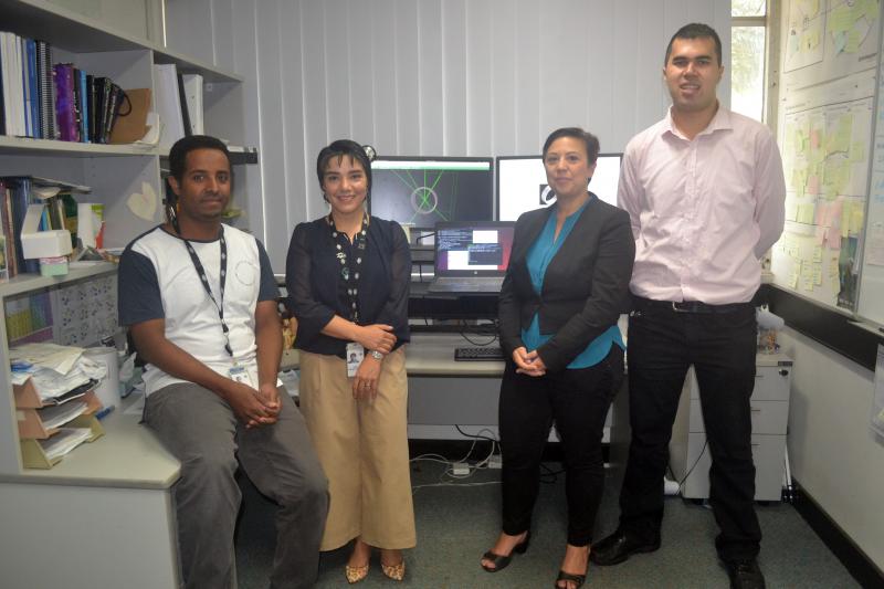
“Once we validate the model, it will be used to generate expected PET images from the treatment plan for comparison to the image acquired during treatment. This will assist with our group’s efforts in developing methods for directly estimating the delivered dose from the measured positron annihilation map,” said Dr Abdella M Ahmed, the primary developer of the software application.
Ahmed is a joint post-doctoral fellow in Human Health at ANSTO and the Centre for Medical Radiation Physics (CMRP) at UOW and his research focuses on modelling of positron emission tomography (PET) systems and developing image reconstruction algorithms.
The Geant4 model was developed under the supervision of Dr Mitra Safavi-Naeini at ANSTO in collaboration with Assoc Prof Susanna Guatelli at the CMRP, Coordinator of the Geant4 Advanced Examples Working Group and was released for the first time within Geant4 10.5 in December 2018.
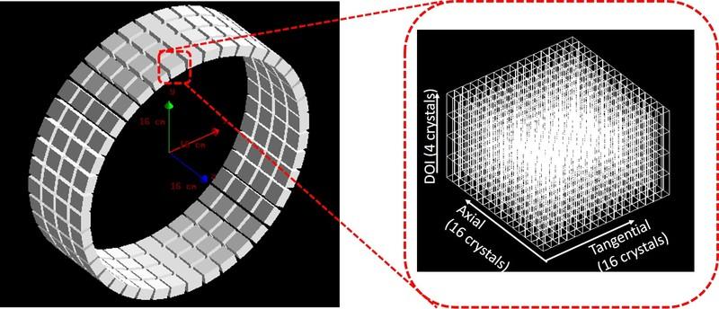
The model is based on a DOI-enabled PET scanner that was developed by Prof Taiga Yamaya and his Imaging Physics team of researchers at the NIRS. In addition to the simulation of the PET scanner, the model includes various types of standard quality assurance devices which are used to compare the performance of PET systems.
“The advanced simulation model will help those who work in the field of nuclear imaging and its applications to particle therapy,” said Safavi-Naeini.
Guatelli added, “It is important that ANSTO contributes to the development of the Geant4 Advanced Examples, which support Geant4 users at international level in the development of specific simulation applications“.
Ahmed, Guatelli and Safavi-Naeini will extend the code to simulate in-beam PET systems used for quality assurance in proton and heavy ion therapy.