
FAQs - Macromolecular Crystallography
Frequently Asked Questions on the Macromolecular Crystallography beamlines (MX1 and MX2)


Showing 61 - 80 of 181 results
Frequently Asked Questions on the Macromolecular Crystallography beamlines (MX1 and MX2)

The Australian crab spider Saccodomus formivorus is the only spider known to weave a unique basket-like web; however, the key to its remarkable design and robust structure is unknown.
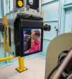
The High Performance Macromolecular Crystallography beamline will enable the study of very small (sub-5 micrometre) or weakly diffracting crystals, providing a state-of-the-art high-throughput facility for researchers. MX3 will be able to study the structures of large proteins and protein complexes for virology, drug design and industrial applications via goniometer mounted crystals, in-tray screening, or via serial crystallography methods.
Monash University, University of Queensland and Australian National University researchers have used ANSTO’s Australian Synchrotron in their study of meteorites found on Earth that could be used in future to find evidence of life on the planet Mars.

In Australia and the Southeast Asia basin, the ANSTO facility offers a wide range of unique nuclear-beam techniques for cultural heritage research.

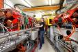
The Accelerator Science group purse a broad research program with the aims of improving the performance and reliability of our accelerators, increasing their research capabilities and developing the next generation of accelerator technology.
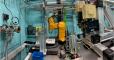
With enhanced submicron spatial resolution, speed and contrast, the Micro-Computed Tomography beamline opens a window on the micron-scale 3D structure of a wide range of samples relevant to many areas of science including life sciences, materials engineering, anthropology, palaeontology and geology. MCT will be able to undertake high-speed and high-throughput studies, as well as provide a range of phase-contrast imaging modalities.

A cross-disciplinary team has used laboratory-based and synchrotron-based infrared spectroscopy imaging techniques to monitor the waxy surface of living plant leaves in real-time to gain insights into plant physiology in response to disease, biological changes or environmental stress.
New infrared imaging technique reveals molecular orientation of proteins in silk fibres
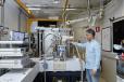
The THz/Far-IR Beamline couples the high brightness and collimation of a bend-magnet synchrotron radiation to a Bruker IFS125HR spectrometer providing high-resolution spectra (0.00096 cm-1) with signal to noise ratio superior to that of thermal sources up to 1350 cm-1 for gas-phase applications; the beamline also delivers signal to noise ratio superior to that of thermal sources up to 350 cm-1 for condensed phase samples.
Hudson Institute of Medical Research and Monash University researchers used synchrotron X-rays produce powerful visualisation of video of changes to blood flow to brain during ventilation in large preterm clinical models.